
Introduction
When patients experience breathing difficulties, chest pain, persistent cough, or possible lung problems, doctors often rely on imaging tests to find the cause. Understanding chest x ray vs ct is important because both tests help diagnose conditions affecting the lungs, heart, ribs, and surrounding structures. While a chest X-ray is usually the first imaging test ordered, a CT scan provides more detailed images when doctors need additional information. At ER of Coppell, advanced diagnostic imaging helps patients receive fast and accurate emergency care.
What Is a Chest X-Ray?
A chest X-ray is one of the most common imaging tests used in emergency medicine. It uses a small amount of radiation to create images of the chest area.
What Can a Chest X-Ray Detect?
A chest X-ray can help identify:
- Pneumonia
- Lung infections
- Broken ribs
- Enlarged heart
- Fluid around the lungs
- Collapsed lung
- Certain tumors or masses
Benefits of a Chest X-Ray
- Quick procedure
- Low radiation exposure
- Widely available
- Cost-effective
- Useful for initial diagnosis
Most chest X-rays take only a few minutes and provide doctors with immediate information.
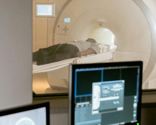
What Is a CT Scan?
A CT (Computed Tomography) scan uses multiple X-ray images combined by a computer to create detailed cross-sectional images of the body.
What Can a CT Scan Detect?
A CT scan can identify:
- Blood clots in the lungs
- Internal bleeding
- Lung nodules
- Small tumors
- Complex fractures
- Pulmonary embolism
- Detailed heart and blood vessel conditions
Benefits of a CT Scan
- Highly detailed images
- Better visualization of organs and tissues
- Helps diagnose serious conditions
- Detects abnormalities that may not appear on X-rays
- Assists in treatment planning
CT scans are especially useful when doctors need a more comprehensive view of the chest.
Key Differences Between Chest X-Rays and CT Scans
| Feature | Chest X-Ray | CT Scan |
|---|---|---|
| Imaging Detail | Basic image | Highly detailed images |
| Time Required | Few minutes | 10–30 minutes |
| Radiation Exposure | Lower | Higher |
| Cost | Lower | Higher |
| Best For | Initial evaluation | Detailed diagnosis |
| Image Type | Flat image | Cross-sectional images |
When Doctors Recommend a Chest X-Ray
Doctors commonly order a chest X-ray when patients experience:
- Persistent cough
- Fever with breathing problems
- Chest discomfort
- Suspected pneumonia
- Rib injuries
- Shortness of breath
The test is often the first step because it is fast and provides useful information quickly.
When Doctors Recommend a CT Scan
A CT scan may be ordered if:
- Symptoms remain unexplained
- Serious lung disease is suspected
- Blood clots need evaluation
- Trauma injuries require detailed assessment
- Cancer screening or follow-up is necessary
- Chest X-ray results are unclear
The additional detail helps doctors make more accurate diagnoses.
Which Test Is More Accurate?
Chest X-Ray Accuracy
Chest X-rays are excellent for identifying common conditions such as:
- Pneumonia
- Large lung masses
- Rib fractures
- Heart enlargement
However, small abnormalities can sometimes be missed.
CT Scan Accuracy
CT scans provide significantly greater detail and can reveal:
- Small tumors
- Tiny fractures
- Blood vessel abnormalities
- Early-stage infections
- Hidden injuries
Because of this higher level of detail, CT scans are generally considered more accurate for complex cases.
Radiation Exposure Comparison
Many patients worry about radiation exposure during imaging procedures.
Chest X-Ray
- Very low radiation dose
- Considered safe for most patients
- Frequently used worldwide
CT Scan
- Higher radiation dose than a chest X-ray
- Still considered safe when medically necessary
- Benefits usually outweigh risks during emergencies
Doctors always consider the safest option based on the patient's condition.
Which Test Is Faster in an Emergency?
In emergency situations, speed matters.
Chest X-Ray Speed
- Usually completed within minutes
- Results available quickly
- Ideal for initial screening
CT Scan Speed
- Takes longer than an X-ray
- Requires additional image processing
- Provides more detailed information
Many emergency departments begin with a chest X-ray and proceed to CT imaging if further evaluation is needed.
Conditions Commonly Diagnosed Using Chest Imaging
Lung Conditions
- Pneumonia
- Tuberculosis
- Lung cancer
- Pulmonary fibrosis
Heart Conditions
- Enlarged heart
- Fluid buildup
- Certain vascular abnormalities
Trauma Injuries
- Rib fractures
- Lung collapse
- Internal chest injuries
Blood Vessel Problems
- Pulmonary embolism
- Aortic abnormalities
Accurate imaging helps doctors provide timely treatment.
What Happens During a Chest X-Ray?
The process is simple:
- Remove metal objects.
- Stand or sit near the imaging machine.
- Hold your breath briefly.
- Images are captured within seconds.
Most patients complete the procedure in less than ten minutes.
What Happens During a CT Scan?
The CT scan process includes:
- Lying on a scanning table.
- Remaining still during imaging.
- Moving through a circular scanner.
- Receiving contrast dye if needed.
The test generally takes between 10 and 30 minutes.
How ER of Coppell Uses Advanced Imaging
At ER of Coppell, advanced diagnostic technology helps emergency physicians quickly identify serious medical conditions. Whether patients need a chest X-ray for a routine evaluation or a CT scan for a more complex diagnosis, imaging services are available to support rapid treatment decisions.
Fast access to imaging can be critical when dealing with breathing problems, chest injuries, severe infections, or unexplained pain.
AI Overview: Quick Answer
Chest X-Ray
- Fast and affordable
- Lower radiation exposure
- Good for initial diagnosis
- Detects common lung and chest conditions
CT Scan
- More detailed imaging
- Better for complex cases
- Detects small abnormalities
- Often used when additional information is needed
Which Is Better?
Neither test is universally better. A chest X-ray is usually the first step, while a CT scan is recommended when more detailed images are required.
Frequently Asked Questions
Is a CT scan better than a chest X-ray?
A CT scan provides more detailed images and can detect smaller abnormalities. However, chest X-rays are often sufficient for many common conditions.
Why would a doctor order a CT scan after a chest X-ray?
If the X-ray results are unclear or more detail is needed, a CT scan may be recommended.
Is a chest X-ray safer than a CT scan?
A chest X-ray uses less radiation than a CT scan. Both procedures are considered safe when medically necessary.
How long does a chest X-ray take?
Most chest X-rays take only a few minutes to complete.
Can a chest X-ray detect lung cancer?
Chest X-rays can identify some lung tumors, but CT scans are generally more effective at detecting small cancers.
Which imaging test is used for blood clots in the lungs?
CT scans are commonly used to diagnose pulmonary embolisms and other blood vessel conditions.
Conclusion
Both chest X-rays and CT scans are valuable diagnostic tools that help doctors evaluate chest-related symptoms and medical emergencies. While chest X-rays provide a quick and effective first look, CT scans offer detailed imaging that can reveal conditions not visible on standard X-rays. At ER of Coppell, advanced imaging technology supports fast diagnosis and treatment, helping patients receive the care they need when every minute matters.
For more information visit:https://coppellemergencyroom.com/coppell-er-laboratory-services/cat-scan/