

Introduction
When a doctor recommends brain imaging, many patients wonder which is better mri or ct scan for brain. The answer is not simply “MRI” or “CT.” Each test has different strengths, and the better option depends on the medical problem being investigated.
A brain MRI uses magnetic fields and radio waves to create highly detailed images without ionizing radiation. CT, or computed tomography, uses X-rays to create cross-sectional images of the brain and skull.
MRI vs. CT Scan for the Brain: Quick Answer
| Feature | Brain MRI | Brain CT |
|---|---|---|
| Image detail | Excellent for soft brain tissue | Good for brain, skull and acute abnormalities |
| Radiation | No ionizing radiation | Uses X-rays |
| Speed | Usually slower | Usually very fast |
| Emergency use | Useful in selected situations | Often preferred initially |
| Head injury | Useful for certain injuries and follow-up | Excellent for bleeding and skull fractures |
| Ischemic stroke | Highly sensitive, especially with diffusion imaging | Fast initial assessment |
| Brain tumors | Excellent soft-tissue detail | Can detect many tumors but usually less detailed |
| Claustrophobia | Can be difficult for some patients | Usually easier |
| Metal implants | Some implants may prevent MRI | Generally compatible |
| Motion sensitivity | More sensitive to movement | Less sensitive to movement |
What Is a Brain MRI?
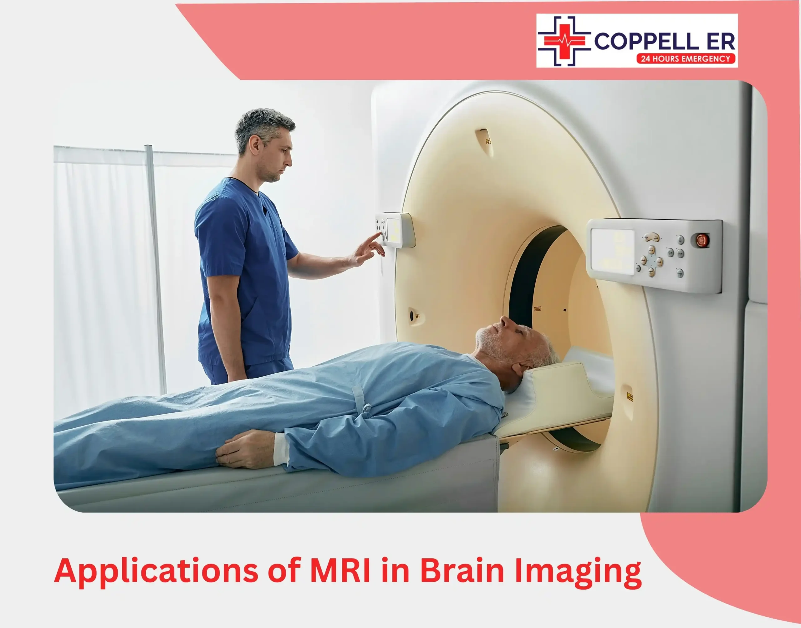
Magnetic resonance imaging creates detailed images using a strong magnetic field, radio waves, and computer processing. Unlike CT, MRI does not use ionizing radiation.
MRI can provide excellent visualization of brain tissue and is commonly used to investigate:
- Brain tumors
- Certain types of stroke
- Multiple sclerosis
- Seizure-related conditions
- Brain infections
- Hydrocephalus
- Certain developmental abnormalities
- Some traumatic brain injuries
- Problems involving the pituitary gland
- Chronic neurological conditions
One important advantage is that MRI can show subtle abnormalities that may be difficult to see on CT.
What Is a Brain CT Scan?
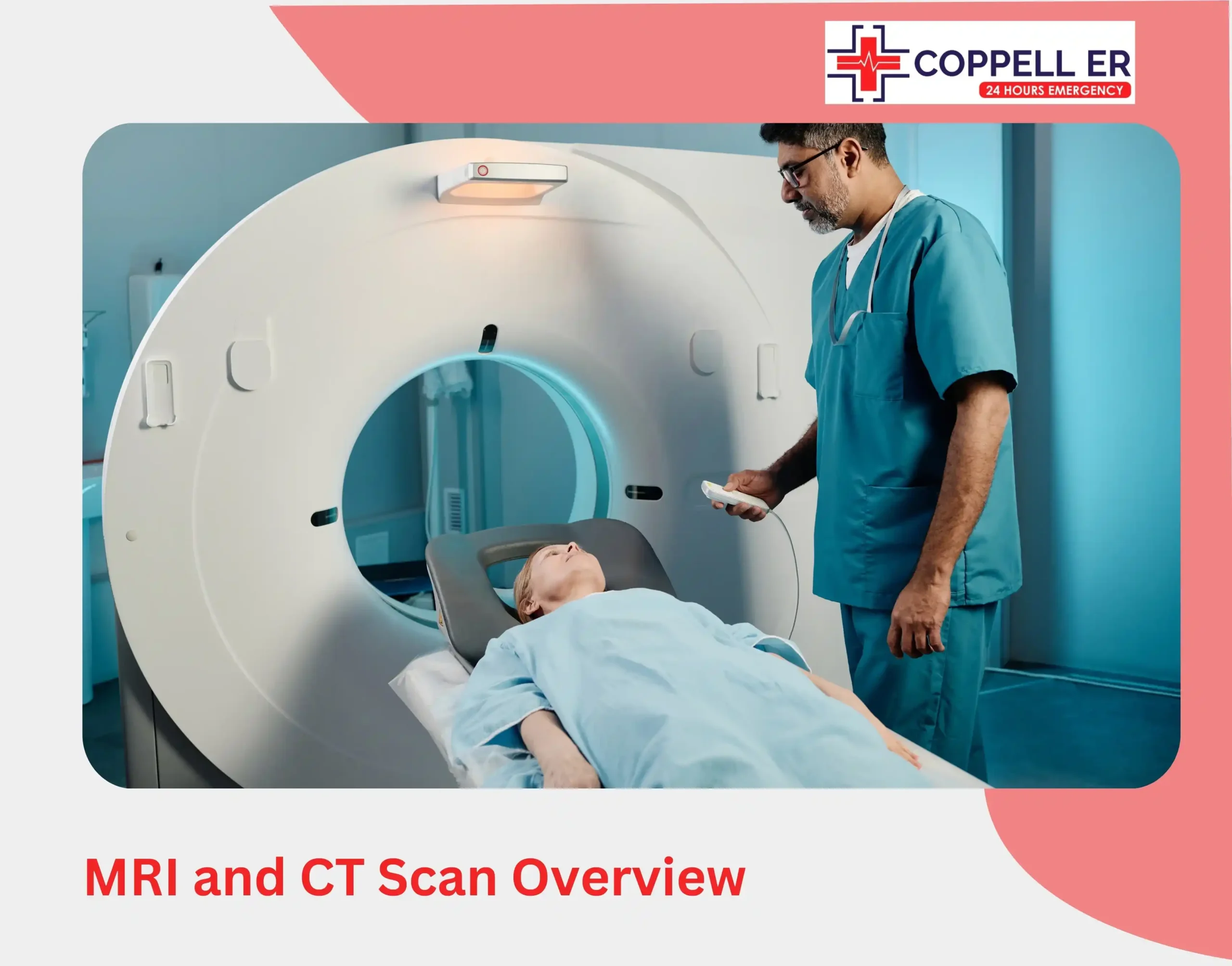
A CT scan uses X-rays and computer processing to create cross-sectional images of the head. It is particularly valuable because the examination can be completed quickly.
Doctors commonly use head CT to evaluate:
- Head injuries
- Suspected brain bleeding
- Skull fractures
- Severe headaches in selected situations
- Certain stroke-related emergencies
- Swelling or pressure inside the skull
- Some tumors
- Certain blood-vessel abnormalities
Why Is CT Better in Many Emergencies?
Speed is one of CT's biggest advantages.
When a person arrives with a serious head injury or possible intracranial bleeding, doctors may need an answer quickly. CT can rapidly show bleeding, swelling and skull fractures.
The American College of Radiology also identifies non-contrast head CT as an appropriate initial test in several acute neurological and head-trauma situations.
MRI or CT: Which Is Better for Different Conditions?
There is no single imaging test that is best for every brain problem.
1. Head Injury
CT is often the first choice in an acute head injury.
It can quickly detect:
- Intracranial bleeding
- Skull fractures
- Brain swelling
- Other serious traumatic abnormalities
MRI may be used later or in selected patients when doctors need additional information about brain tissue or injuries that are not clearly visible on CT.
2. Suspected Stroke
The answer depends on the type of stroke and the urgency.
CT is frequently used rapidly in emergency stroke evaluation, particularly to determine whether bleeding is present. MRI with diffusion-weighted imaging can be more sensitive for early ischemic stroke and small areas of brain injury.
3. Brain Tumors
MRI is generally better for detailed evaluation of brain tumors because it provides excellent soft-tissue contrast.
MRI can help doctors assess the tumor's location, characteristics and relationship to nearby brain structures. CT may also identify tumors and can be useful in certain circumstances, including evaluating bone or when MRI is not appropriate.
4. Brain Bleeding
CT is commonly used when doctors need to quickly identify acute bleeding.
MRI can also identify hemorrhage, but the speed and accessibility of CT make it particularly valuable in emergency settings.
5. Multiple Sclerosis and Other Neurological Disorders
MRI is generally preferred for many conditions involving subtle changes in brain tissue.
Because MRI provides detailed images of soft tissue without ionizing radiation, it is an important tool for evaluating conditions such as multiple sclerosis and other neurological disorders.
FAQs
Is MRI more accurate than CT for the brain?
MRI can be more sensitive than CT for many abnormalities involving brain tissue, particularly subtle or early changes. However, CT can be the better test for urgent conditions such as acute bleeding or certain traumatic injuries.
Is CT or MRI better for a brain tumor?
MRI is generally preferred for detailed evaluation of brain tumors because it provides excellent soft-tissue detail. CT may still be useful in certain circumstances.
Which scan is better for a stroke?
Both can be important. CT is often used rapidly in emergency stroke evaluation, while MRI with diffusion-weighted imaging can detect many ischemic strokes earlier and with greater sensitivity.
Final Takeaway
MRI and CT scans are complementary rather than competing technologies. MRI is usually the stronger choice for detailed brain-tissue imaging, while CT is often the stronger choice when speed, bleeding detection or evaluation of acute trauma is the priority.
For more blogs visit : https://caecusarguo779260.substack.com/p/7-powerful-echocardiogram-facts-you?r=5yu06d&utm_campaign=post&utm_medium=web&showWelcomeOnShare=true

 English
 Arabic
 Spanish
 Portuguese
 Deutsch
 Turkish
 Dutch
 Italiano
 Russian
 Romaian
 Portuguese (Brazil)
 Greek